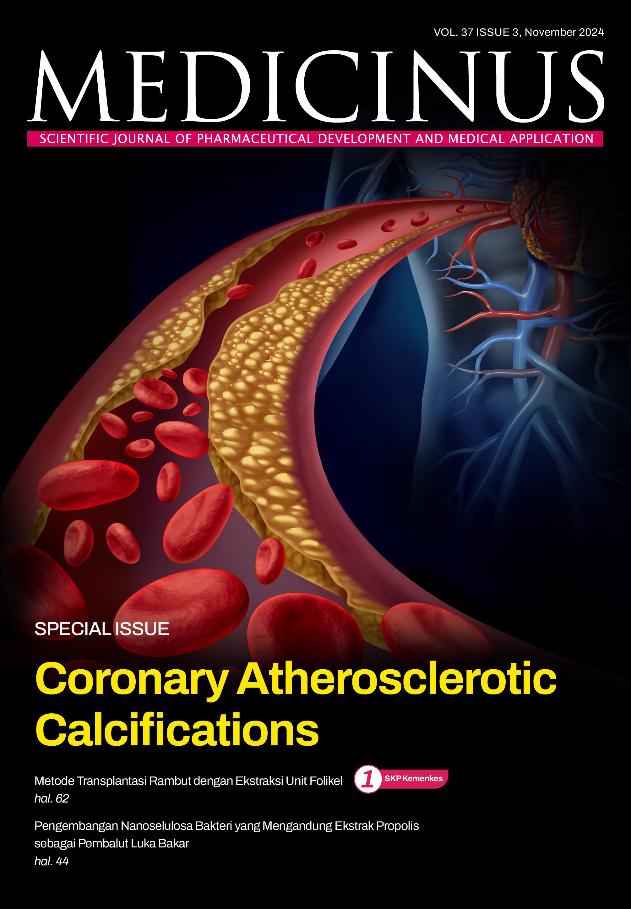
Coronary Atherosclerotic Calcification
DOI:
https://doi.org/10.56951/00pm8d52Kata Kunci:
penyakit arteri koroner, kalsifikasi aterosklerosis, kalsifikasi arteri koronerAbstrak
Penyakit arteri koroner merupakan masalah kesehatan masyarakat yang signifikan di Asia Tenggara, termasuk Indonesia. Penyakit ini ditandai oleh penumpukan plak aterosklerotik di dalam arteri koroner, yang menyebabkan aliran darah ke miokardium berkurang atau terhenti. Salah satu ciri khas aterosklerosis adalah adanya kalsifikasi, yang dapat terjadi selama progresi maupun regresi plak. Hingga saat ini, pola perkembangan lesi aterosklerotik belum sepenuhnya jelas apakah mengikuti urutan dengan pola linear. Pembentukan mikrokalsifikasi memulai proses kalsifikasi dan memerlukan
penelitian lebih lanjut. Beberapa modalitas pencitraan yang telah berkembang pesat dapat membantu mengukur beban kalsifikasi arteri koroner dan memandu manajemen yang tepat. Isu tentang paparan radiasi telah menjadikan pencitraan kalsium arteri koroner sebagai parameter yang paling efektif, efisien, dan mudah diamati di antara teknik pencitraan invasif dan non-invasif. Mengidentifikasi karakteristik plak berisiko tinggi serta kalsifikasi yang berat atau ekstensif sangat penting untuk memberikan tata laksana yang optimal. Namun, perkembangan kalsifikasi masih belum pasti, dan penelitian terus
dilakukan untuk mengeksplorasi metode potensial yang dapat membalikkan proses ini. Berbagai alat dan teknik kompleks telah dikembangkan untuk menangani berbagai jenis lesi yang mengalami kalsifikasi. Penelitian lebih lanjut diperlukan untuk mengidentifikasi pasien yang mengalami perkembangan kalsifikasi yang lebih cepat, untuk mengoptimalkan strategi pencegahan.
Unduhan
Referensi
Mensah GA, Fuster V, Murray CJL, Roth GA, Mensah GA, Abate YH, et al. Global burden of cardiovascular
diseases and risks 1990-2022. J Am Coll Cardiol. 2023;82(25):2350–473.
Ralapanawa U, Sivakanesan R. Epidemiology and the magnitude of coronary artery disease and acute coronary syndrome: a narrative review. J Epidemiol Glob Health 2021;11(2):169. DOI: https://doi.org/10.2991/jegh.k.201217.001
Badan Penelitian dan Pengembangan Kesehatan Kementerian Kesehatan Republik Indonesia. Laporan Nasional Riskesdas 2018.
Adisasmito W, Amir V, Atin A, Megraini A, Kusuma D. Geographic and socioeconomic disparity in cardiovascular risk factors in Indonesia: analysis of the basic health research 2018. BMC Public Health 2020;20(1):1004. DOI: https://doi.org/10.1186/s12889-020-09099-1
Jadhav KP, Kavalipatu KNR, Kuchulakanti PK, Reddy RP, Athuluri R, Prakash G, et al. Coronary artery calcification: from cell to stent—a review. Indian Journal of Clinical Cardiology 2021;2(2):97–109. DOI: https://doi.org/10.1177/26324636211013156
Mori H, Torii S, Kutyna M, Sakamoto A, Finn A V., Virmani R. Coronary artery calcification and its progression: what does it really mean? JACC Cardiovasc Imaging 2018;11(1):127–42. DOI: https://doi.org/10.1016/j.jcmg.2017.10.012
Onnis C, Virmani R, Kawai K, Nardi V, Lerman A, Cademartiri F, et al. Coronary artery calcification: current concepts and clinical implications. Circulation 2024;149(3):251-66. DOI: https://doi.org/10.1161/CIRCULATIONAHA.123.065657
Parikh P, Shah N, Ahmed H, Schoenhagen P, Fares M. Coronary artery calcium scoring: its practicality and clinical utility in primary care. Cleve Clin J Med. 2018;85(9):707. DOI: https://doi.org/10.3949/ccjm.85a.17097
Virmani R, Kolodgie FD, Burke AP, Farb A, Schwartz SM. Lessons from sudden coronary death: a comprehensive morphological classification scheme for atherosclerotic lesions. Arterioscler Thromb Vasc Biol. 2000;20(5):1262-75. DOI: https://doi.org/10.1161/01.ATV.20.5.1262
Nakahara T, Dweck MR, Narula N, Pisapia D, Narula J, Strauss HW. Coronary artery calcification: from mechanism to molecular imaging. JACC Cardiovasc Imaging. 2017;10(5):582–93. DOI: https://doi.org/10.1016/j.jcmg.2017.03.005
Nguyen PTH, Coche E, Goffin E, Beguin C, Vlassenbroek A, Devuyst O, et al. Prevalence and determinants of coronary and aortic calcifications assessed by chest CT in renal transplant recipients. Am J Nephrol. 2007;27(4):329–35. DOI: https://doi.org/10.1159/000102978
Pezel T, Sideris G, Dillinger JG, Logeart D, Manzo-Silberman S, Cohen-Solal A, et al. Coronary computed tomography angiography analysis of calcium content to identify non-culprit vulnerable plaques in patients with acute coronary syndrome. Front Cardiovasc Med. 2022;15;9. DOI: https://doi.org/10.3389/fcvm.2022.876730
Lanzer P, Hannan FM, Lanzer JD, Janzen J, Raggi P, Furniss D, et al. Medial arterial calcification: JACC state-of-the-art review. J Am Coll Cardiol. 2021;78(11):1145-65. DOI: https://doi.org/10.1016/j.jacc.2021.06.049
Shreya D, Zamora DI, Patel GS, Grossman I, Rodriguez K, Soni M, et al. Coronary artery calcium score - a reliable indicator of coronary artery disease? Cureus. 2021;13(12):e20149. DOI: https://doi.org/10.7759/cureus.20149
Hecth HS, Blaha MJ, Kazerooni EA, Cury RC, Budoff M, Leipsic J, et al. Journal of cardiovascular computed tomography CAC-DRS: coronary artery calcium data and reporting system. An expert consensus document of the Society of Cardiovascular Computed Tomography (SCCT). J Cardiovasc Comput Tomogr. 2018;12(3):185-91. DOI: https://doi.org/10.1016/j.jcct.2018.03.008
Vancheri F, Longo G, Vancheri S, Danial JSH, Henein MY. Coronary artery microcalcification: imaging and clinical implications. Diagnostics 2019;9(4):1-17. DOI: https://doi.org/10.3390/diagnostics9040125
Nicoll R, Henein M. Arterial calcification: a new perspective? Int J Cardiol. 2017;228:11-22. DOI: https://doi.org/10.1016/j.ijcard.2016.11.099
Sharma RK, Voelker DJ, Sharma RK, Singh VN, Bhatt G, Moazazi M, et al. Coronary computed tomographic angiography (CCTA) in community hospitals: “current and emerging role”. Vasc Health Risk Manag 2010;6:307-16. DOI: https://doi.org/10.2147/VHRM.S9108
Tzolos E, Dweck MR. 18F-Sodium Fluoride (18F-NaF) for imaging microcalcification Activity in the cardiovascular ;system. Arterioscler Thromb Vasc Biol. 2020;40(7):1620–6. DOI: https://doi.org/10.1161/ATVBAHA.120.313785
McInerney A, Escaned J, Gonzalo N. Calcified coronary artery disease: pathophysiology, intracoronary imaging assessment, and plaque modification techniques. REC Interv Cardiol. 2022;4(3):216-27. DOI: https://doi.org/10.24875/RECICE.M22000291
Cury RC, Blankstein R, Leipsic J, Abbara S, Achenbach S, Berman D, et al. CAD-RADSTM 2.0 - 2022 coronary artery disease – reporting and data system an expert consensus document of the Society of Cardiovascular Computed Tomography (SCCT), the American College of Cardiology (ACC), the American College of Radiology (ACR) and the North America society of cardiovascular imaging (NASCI). J Cardiovasc Comput Tomogr. 2022;16(6):536-57. DOI: https://doi.org/10.1148/ryct.220183
Maron DJ, Budoff MJ, Sky JC, Bommer WJ, Epstein SD, Fisher DA, et al. Coronary artery calcium staging to guide preventive interventions: a proposal and call to action. JACC Adv. 2024;3(11):101287. DOI: https://doi.org/10.1016/j.jacadv.2024.101287
Budoff MJ, Kinninger A, Gransar H, Achenbach S, Al-Mallah M, Bax JJ, et al. When does a calcium score equate to secondary prevention?: insights from the multinational CONFIRM Registry. JACC Cardiovasc Imaging. 2023;16(9):1181–9. DOI: https://doi.org/10.1016/j.jcmg.2023.03.008
Marx, N., Federici, M., Schütt, K., Müller-Wieland, D., Ajjan, R. A., Antunes, et al. 2023 ESC Guidelines for the management of cardiovascular disease in patients with diabetes. European Heart Journal 2023;44(39):4043–140. DOI: https://doi.org/10.1093/eurheartj/ehad192
Budoff MJ, Bhatt DL, Kinninger A, Lakshmanan S, Muhlestein JB, Le VT, et al. Effect of icosapent ethyl on progression of coronary atherosclerosis in patients with elevated triglycerides on statin therapy: final results of the EVAPORATE trial. European Heart Journal 2020;41:3925–32. DOI: https://doi.org/10.1093/eurheartj/ehaa652
Golub IS, Misic A, Schroeder LP, Aldana-Bitar J, Krishnan S, Kianoush S, et al. Calcific coronary lesions: management, challenges, and a comprehensive review. AIMS Med Sci 2024;11(3):292–317. DOI: https://doi.org/10.3934/medsci.2024021
Hollenberg EJ, Lin F, Blaha MJ, Budoff MJ, van den Hoogen IJ, Gianni U, et al. Relationship between coronary artery calcium and atherosclerosis progression among patients with suspected coronary artery disease. JACC: Cardiovascular Imaging 2022;15(6):1063-74. DOI: https://doi.org/10.1016/j.jcmg.2021.12.015
Ali ZA, Galougahi KK, Mintz GS, Maehara A, Shlofmitz RA, Mattesini A. Intracoronary optical coherence tomography: state of the art and future directions. EuroIntervention 2021;17(2):e105-23. DOI: https://doi.org/10.4244/EIJ-D-21-00089
Unduhan
Terbitan
Bagian
Diterbitkan
Unduhan
Lisensi
Hak Cipta (c) 2024 Sony Hilal Wicaksono, Christian Setiawan, Indah Fitriani

Artikel ini berlisensi Creative Commons Attribution-NonCommercial 4.0 International License.
Cara Mengutip


