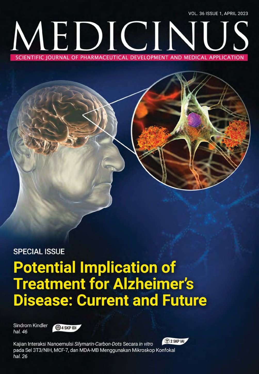
Potential Implication of Treatments for Alzheimer’s Disease: Current and Future
DOI:
https://doi.org/10.56951/medicinus.v36i1.112Kata Kunci:
penyakit Alzheimer, β-amyloid, neurofibrillary tangles, gangguan visual, proses inflamasi, pengobatan herbalAbstrak
Penyakit Alzheimer (AD) adalah penyakit neurodegeneratif yang progresif, penyebab mayoritas kasus demensia, yang ditandai dengan kelainan struktural anatomi otak. Penderita AD kehilangan siklus normal aktivitas sehari-hari, yang secara bertahap akan memperburuk ingatan dan keterampilan berkomunikasi, sering pula disertai dengan gangguan penglihatan, yang berlanjut ke arah kondisi disabilitas. Karakteristik patologis otak penderita AD adalah terjadinya plak β-amyloid (Aβ) dan terbentuknya neurofibrillary tangles. Terapi yang ada hingga saat ini hanya menargetkan untuk membantu menurunkan gejala dengan menggunakan berbagai obat dan psikoterapi, akan tetapi tidak menyembuhkan penyakit. Beberapa obat kimiawi untuk aplikasi klinis sejauh ini hanya mencapai efek terapeutik untuk tujuan pencegahan. Pengobatan tradisional herbal selama ribuan tahun telah mengumpulkan banyak pengalaman dalam terapi demensia. Studi farmakologi modern mengonfirmasi efek terapeutik dari banyak komponen aktif obat herbal. Efek terapeutik dari obat herbal ini dinilai sangat potensial untuk berbagai penyakit, termasuk gangguan neurodegeneratif.
Unduhan
Referensi
Scheltens NM, Galindo-Garre F, Pijnenburg YA, et al. The identification of cognitive subtypes in Alzheimer’s disease dementia using latent class analysis. J Neurol Neurosurg Psychiatry 2015;87(3):235–43. DOI: https://doi.org/10.1136/jnnp-2014-309582
Graeber MB, Kosel S, Egensperger R, et al. Rediscovery of the case described by Alois Alzheimer in 1911: historical, histological and molecular genetic analysis. Neurogenetics 1997;1(1):73–80. DOI: https://doi.org/10.1007/s100480050011
Petersen RC, Smith GE, Kokmen E. Mild cognitive impairment. Clinical characterization and outcome. Arch Neurol 1999;46:303-8. DOI: https://doi.org/10.1001/archneur.56.3.303
Palmer K, Berger AK, Monastero R, Windblad B, Backman L, Fratilioni L. Predictors of progression from mild cognitive impairment to Alzheimer disease. Neurology 2007;68:1596-602. DOI: https://doi.org/10.1212/01.wnl.0000260968.92345.3f
Wolf H, Grundwald M, Ecke GM, et al. The prognosis to mild cognitive impairment in the elderly. J Neural Transm Supp 1998;54:31-50. DOI: https://doi.org/10.1007/978-3-7091-7508-8_4
Lopez OL, Becker JT, Sweer RA. Non-cognitive symptoms in mild cognitive impairment subjects. Neurocase 2005;11:65-71. DOI: https://doi.org/10.1080/13554790490896893
Cumming JL. Behavioral and neuropsychiatric outcomes in Alzheimer’ disease. CNS Spectr 2005;10(Supp18):22-5. DOI: https://doi.org/10.1017/S1092852900014206
Alipour F, Mohammadzadeh E, Khallaghi B. Evaluation of apoptosis in rat hippocampal tissue in an experimental model of alzheimer’s disease. Neurosci J Shefaye Khatam 2014;22:13–20. DOI: https://doi.org/10.18869/acadpub.shefa.2.2.13
Kaeser P-F, Ghika J, Borruat F-X. Visual signs and symptoms in patients with the visual variant of Alzheimer disease. BMC Ophthalmol 2015;15:65. DOI: https://doi.org/10.1186/s12886-015-0060-9
Gao S, Hendrie HC, Hall KS, Hui S. The relationships between age, sex, and the incidence of dementia and Alzheimer disease: a meta-analysis. Arch Gen Psychiatry 1998;55:809-15. DOI: https://doi.org/10.1001/archpsyc.55.9.809
Prince M, Wimo A, Guerchet M, et al. World Alzheimer Report 2015—The global impact of dementia: An analysis of prevalence, incidence, cost and trends. London: Alzheimer’s Disease International, 2015.
Brookmeyer R, Johnson D, Ziegler-Graham K, Arrigh HM. Forecasting the global burden of Alzheimer's disease. Alz Dementia 2007;3:186-91. DOI: https://doi.org/10.1016/j.jalz.2007.04.381
Klein BEK, Moss SE, Klein R, Lee KE, Cruickshanks KJ. Associations of visual function with physical outcomes and limitations 5 years later in an older population: The Beaver Dam eye study. Ophthalmology 2003;110:644–50. DOI: https://doi.org/10.1016/S0161-6420(02)01935-8
Jindal H, Bhatt B, Sk S, Singh Malik J. Alzheimer disease immunotherapeutics: then and now. Human vaccines immunotherapeutics 2014;10(9):2741–3. DOI: https://doi.org/10.4161/21645515.2014.970959
Tobinick E, Gross H, Weinberger A, Cohen H. TNF-alpha Modulation for Treatment of Alzheimer's Disease: A 6-Month Pilot Study. Medscape Gen Medicine 2006;8(2):25. DOI: https://doi.org/10.1016/j.jalz.2006.05.1244
Tan ZS, Beiser AS, Vasan RS, et al. Inflammatory markers and the risk of Alzheimer disease: the Framingham Study. Neurology 2008;70:1222-3. DOI: https://doi.org/10.1212/01.wnl.0000307660.86647.7b
McEwen BS. Effects of adverse experiences for brain structure and function. Biol Psychiatry 2000;48:721-31. DOI: https://doi.org/10.1016/S0006-3223(00)00964-1
Mullan M. Familial Alzheimer's disease: second gene locus located. BMJ 1992;305:1108-9. DOI: https://doi.org/10.1136/bmj.305.6862.1108
Schellenberg GD, Boehnke M, Wijsman EM, et al. Genetic association and linkage analysis of the locus and familial Alzheimer's disease. Ann Neurol 1992;31:223-7. DOI: https://doi.org/10.1002/ana.410310214
Poirier J, Davignon J, Bouthillier D, Kogan S, Bertrand P, Gauthier S. Apolipoprotein E polymorphism and Alzheimer’s disease. Lancet 1993;342:697–9. DOI: https://doi.org/10.1016/0140-6736(93)91705-Q
Karen S, Tim W, Karoline K, Oliver S, Tim S, Zuzana W. Rate of Cognitive Decline in Alzheimer’s Disease Stratified by Age. J Alzheimer's Dis 2019;69:1153-60. DOI: https://doi.org/10.3233/JAD-181047
Popovic N, Brundin P. Therapeutic potential of controlled drug delivery systems in neurodegenerative diseases. Int J Pharm 2006;314:120–6. DOI: https://doi.org/10.1016/j.ijpharm.2005.09.040
Gosche KM, Mortimer JA, Smith CD, Markesbery WR, Snowdon DA. Hippocampal volume as an index of Alzheimer neuropathology: findings from the Nun Study. Neurology 2002;58:1476–82. DOI: https://doi.org/10.1212/WNL.58.10.1476
Vemuri P, Wiste HJ, Weigand SD, et al. MRI and CSF biomarkers in normal, MCI, and AD subjects: diagnostic discrimination and cognitive correlations. Neurology 2009;73:287–93. DOI: https://doi.org/10.1212/WNL.0b013e3181af79e5
Hua X, Leow AD, Parikshak N, et al.Tensor-based morphometry as a neuroimagingbiomarker for Alzheimer’s disease: an MRI study of 676 AD, MCI, and normal subjects. Neuroimage 2008;43:458–69. DOI: https://doi.org/10.1016/j.neuroimage.2008.07.013
Braak H, Braak E, Bohl J. Staging of Alzheimer-related cortical destruction. Eur Neurol 1993;33:403–8. DOI: https://doi.org/10.1159/000116984
Heinonen O, Soininen H, Sorvari H, Kosunen O, Paljärvi L, Koivisto E. Loss of synaptophysin-like immunoreactivity in the hippocampal formation is an early phenomenon in Alzheimer's disease. Neuroscience 1995;64:375–84. DOI: https://doi.org/10.1016/0306-4522(94)00422-2
Koffie RM, Meyer-Luehmann M, Hashimoto T. Oligomeric amyloid beta associates with postsynaptic densities and correlates with excitatory synapse loss near senile plaques. Proceed Nat Acad Sci 2009;106:4012-7. DOI: https://doi.org/10.1073/pnas.0811698106
López-Hernández GY, Thinschmidt JS, Morain P, et al. Positive modulation of alpha7- nAChR responses in rat hippocampal interneurons to full agonists and the alpha-selective partial agents, 40H-GTS-21 and S 24795. Neuropharmacology 2009;56:821-30. DOI: https://doi.org/10.1016/j.neuropharm.2009.01.011
Kook, SH. Seok Hong, M. Moon, and I. Mook-Jung. Disruption of blood-brain barrier in Alzheimer disease pathogenesis, Tissue Barriers 2014;1(2):e23993. DOI: https://doi.org/10.4161/tisb.23993
Salminen A, Ojala J, Kauppinen A, Kaarniranta K, Suuronen T. Inflammation in Alzheimer's disease: amyloid-beta oligomers trigger innate immunity defence via pattern recognition receptors. Prog Neurobiol 2009;87:181-94. DOI: https://doi.org/10.1016/j.pneurobio.2009.01.001
Selkoe DJ. Alzheimer's disease: A central role for amyloid. J Neuropathol Exp Neurol 1994;53:438–47. DOI: https://doi.org/10.1097/00005072-199409000-00003
Hardy J, Selkoe DJ. The amyloid hypothesis of Alzheimer's disease: progress and problems on the road to therapeutics. Science 2002;297:353–6. DOI: https://doi.org/10.1126/science.1072994
Abramov E, Dolev I, Fogel H, Ciccotosto GD, Ruff E and Slutsky I. Amyloid β as a positive endogenous regulator of release probability at hippocampal synapses, Nat Neurosci 2009;12:1567-76. DOI: https://doi.org/10.1038/nn.2433
Panza F, Solfrizzi V, Frisardi V, et al. Beyond the neurotransmitter-focused approach in treating Alzheimer's disease: drugs targeting beta-amyloid and tau protein. Aging Clin Exp Res 2009;21:386-406. DOI: https://doi.org/10.1007/BF03327445
Querfurth HW, LaFerla FM. Alzheimer's disease. N Engl J Med 2010;362:329-44. DOI: https://doi.org/10.1056/NEJMra0909142
Graeber MB. Changing face of microglia. Science 2010;330:783-8. DOI: https://doi.org/10.1126/science.1190929
Fuhrmann M, Bittner T, Jung CK, et al. Microglial Cx3cr1 knockout prevents neuron loss in a mouse model of Alzheimer's disease. Nat Neurosci 2010;13:411-3. DOI: https://doi.org/10.1038/nn.2511
Tahara K, Kim HD, Jin JJ, Maxwell JA, Li L, Fukuchi K. Role of toll-like receptor signalling in Abeta uptake and clearance. Brain 2006;129:3006-19. DOI: https://doi.org/10.1093/brain/awl249
Walker, Douglas; Lue, Lih-Fen. Anti-inflammatory and Immune Therapy for Alzheimer's Disease: Current Status and Future Directions: Current Neuropharmacology 2007;5: 232-43. DOI: https://doi.org/10.2174/157015907782793667
Gilmore GC, Whitehouse PJ. Contrast sensitivity in Alzheimer’s disease: a 1-year longitudinal analysis. Optom Vis Sci 1995;72:83–91. DOI: https://doi.org/10.1097/00006324-199502000-00007
Mendez MF, Cherrier MM, Meadows RS. Depth perception in Alzheimer’s disease. Percept Mot Skills 1996;83:987–995. DOI: https://doi.org/10.2466/pms.1996.83.3.987
Trick GL, Trick LR, Morris P, Wolf M. Visual field loss in senile dementia of the Alzheimer’s type. Neurology 1995;45:68-74. DOI: https://doi.org/10.1212/WNL.45.1.68
Rizzo JF 3rd1, Cronin-Golomb A, Growdon JH, et al. Retinocalcarine function in Alzheimer’s disease: a clinical and electrophysiological study. Arch Neurol 1992;49:93-101. DOI: https://doi.org/10.1001/archneur.1992.00530250097023
Armstrong RA. Visual field defects in Alzheimer’s disease patients may reflect differential pathology in the primary visual cortex. Optom Vis Sci 1996;73:677–82. DOI: https://doi.org/10.1097/00006324-199611000-00001
Victoria S Pelak and William Hills. Vision in Alzheimer's Disease: A Focus on the Anterior Afferent Pathway. Neurodegener Dis Manag 2018;8:49-67. DOI: https://doi.org/10.2217/nmt-2017-0030
Goldstein LE, Muffat JA, Cherny RA. Cytosolic beta-amyloid deposition and supranuclear cataracts in lenses from people with Alzheimer's disease. Lancet 2003;361:1258-65. DOI: https://doi.org/10.1016/S0140-6736(03)12981-9
Berisha F, Feke GT, Trempe CL, McMeel JW, Schepens CL. Retinal Abnormalities in Early Alzheimer’s Disease. Invest Ophthal Visual Science 2007;48:2285-9. DOI: https://doi.org/10.1167/iovs.06-1029
Donnelly RJ, Friedhoff AJ, Beer B, Blume AJ, Vitek MP. Interleukin-1 stimulates the beta-amyloid precursor protein promoter. Cell Mol Neurobiol 1990;10:485-95. DOI: https://doi.org/10.1007/BF00712843
Kawas CH, Corrada MM, Brookmeyer R, et al. Visual memory predicts Alzheimer's disease more than a decade before diagnosis. Neurology 2003;60:1089-93. DOI: https://doi.org/10.1212/01.WNL.0000055813.36504.BF
Hinton DR, Sadun AA, Blanks JC, Miller CA. Optic-nerve degeneration in Alzheimer’s disease. N Engl J Med 1986;315:485–7. DOI: https://doi.org/10.1056/NEJM198608213150804
Wostyn P, K Audenaert K, De Deyn PP. Alzheimer’s disease and glaucoma: Is there a causal relationship? Br J Ophthalmol 2009;93:1557-9 . DOI: https://doi.org/10.1136/bjo.2008.148064
Zhu X-C, Yu Y, Wang H-F, et al. Physiotherapy intervention in Alzheimer's disease: systematic review and meta-analysis. J Alzheimers Dis 2015;44:163-74. DOI: https://doi.org/10.3233/JAD-141377
Coley N, Gallini A, Andrieu S. Prevention studies in Alzheimer’s disease: progress towards the development of new therapeutics. CNS Drugs 2015;29:519-28. DOI: https://doi.org/10.1007/s40263-015-0256-9
Zhang L, Liu JJ, Zhao Y, Liu Y, Lin JW. N-butylphthalide affects cognitive function of APP/PS1 transgenic mice (Alzheimer’s disease model). Zhongguo Zuzhi Gongcheng Yanjiu 2019;23:3025-30.
Shahaji FA, Chavan Sadhana P. A Review on Alzheimer’s disease and its concepts in Ayurveda. Internat J Ayurveda and Pharma Res 2015;3:52-6.
Zenaro E, Piacentino G, and Constantin G. The blood-brain barrier in Alzheimer's disease. Neurobiol Dis 2017;107:41–56. DOI: https://doi.org/10.1016/j.nbd.2016.07.007
Mishra S, Palanivelu K. The effect of curcumin (turmeric) on Alzheimer’s disease: An overview. Ann Indian Acad Neurol 2008;11:13-19. DOI: https://doi.org/10.4103/0972-2327.40220
Roy A. Role of medicinal plants against Alzheimer’s disease. Int J Complement Alt Med. 2018;11:205‒208. DOI: https://doi.org/10.15406/ijcam.2018.11.00398
Kwoka BHB, Koha B, Ndubuisia MI, Elofssona M, Crewsa CM. The anti-inflammatory natural product parthenolide from themedicinal herb Feverfew directly binds to and inhibits IUB kinase. Chem Biol 2001;8:759-66. DOI: https://doi.org/10.1016/S1074-5521(01)00049-7
Phani Kumar G and Khanum F. Neuroprotective potential of phytochemicals Pharmacog Rev 2012;6:81–90. DOI: https://doi.org/10.4103/0973-7847.99898
Chen Y X, Wu S, Yu X, et al. Neuroprotection of tanshinone IIA against cerebral ischemia/reperfusion injury through inhibition of macrophage migration inhibitory factor in rats. Plos One 2012;7:e40165. DOI: https://doi.org/10.1371/journal.pone.0040165
Li J, Fang-Yin F-X, Yuan Y. Pharmacokinetics of phenolic compounds of Danshen extract in rat blood and brain by microdialysis sampling. J Ethnopharmacol 2011;136:129-36. DOI: https://doi.org/10.1016/j.jep.2011.04.023
Wang R, Li YN, Wang GJ, Hao HE, Wu XL and Zhou F. Neuroprotective effects and brain transport of Ginsenoside Rg1. Chin J Nat Med 2009;7:315–20. DOI: https://doi.org/10.3724/SP.J.1009.2008.00315
Wu CF, LiBi X, YuYang J, et al. Differential effects of ginsenosides on NO and TNF-α production by LPS-activated N9 microglia. International Immunopharmacol 2007;7:313-20. DOI: https://doi.org/10.1016/j.intimp.2006.04.021
Hook V, Toneff T, Bogyo M, et al. Inhibition of cathepsin B reduces β-amyloid production in regulated secretory vesicles of neuronal chromaffin cells: Evidence for cathepsin B as a candidate β-secretase of Alzheimer’s disease. Biol Chem 2005;386:931–40. DOI: https://doi.org/10.1515/BC.2005.108
Mueller-Steiner S, Zhou Y, Arai H, et al. Antiamyloidogenic and neuroprotective functions of cathepsin B: Implications for Alzheimer’s disease. Neuron 2006;51:703–14. DOI: https://doi.org/10.1016/j.neuron.2006.07.027
Pocernich CB and Butterfield DA. Elevation of Glutathione as a Therapeutic Strategy in Alzheimer Disease. Biochim Biophys Acta 2012;1822(5):625–30. DOI: https://doi.org/10.1016/j.bbadis.2011.10.003
Liu X, Zhang Y, Yang X (2019) PM2.5 induced neurodegenerative-like changes in mice and the antagonistic effects of vitamin E. Toxicol Res 2019;8:172– 9. DOI: https://doi.org/10.1039/C8TX00333E
Shi X, Zheng Z, Li J, et al. Curcumin inhibits Aβ-induced microglial inflammatory responses in vitro: Involvement of ERK1/2 and p38 signaling pathways Neurosci Lett 2015;594:105-10. DOI: https://doi.org/10.1016/j.neulet.2015.03.045
Salehi B , Calina D, Docea AO, et al. Curcumin's Nanomedicine Formulations for Therapeutic Application in Neurological Diseases. J Clin Med Actions 2020;9:430. DOI: https://doi.org/10.3390/jcm9020430
Unduhan
Terbitan
Bagian
Diterbitkan
Unduhan
Lisensi
Hak Cipta (c) 2023 Jan Sudir Purba

Artikel ini berlisensi Creative Commons Attribution-NonCommercial 4.0 International License.


